Annual Oration 1960
By Max Ritvo, M.D.
The Annual Oration or Discourse has become a time-honored custom. I am deeply appreciative of the honor of having been elected to deliver it, particularly since it is but the second time that a roentgenologist has had this great privilege.
Every physician is interested in the x-ray because it is utilized almost universally in the diagnosis of human ills. I was influenced in my choice of subject by the fact that many members of the Society were among the first to appreciate the significance of Roentgen's contribution, with the result that some of the most important applications of the rays as a practical medical tool originated in Boston soon after the announcement of their discovery.
On November 8, 1895, Wilhelm Conrad Roentgen first saw a "light" on a fluorescent screen that had never before been observed by the human eye. He submitted a paper entitled "On a New Kind of Ray" to the Physical Medical Society of Würzburg on December 28. The news soon became public, and the report of a ray "which for purposes of photography will penetrate wood, cloth and most other solid substances" created great excitement and enthusiasm. Scientists everywhere immediately began to work with the rays. The Boston Medical and Surgical Journal of February 13, 1896, carried a reproduction of a roentgenogram of a human hand.
A pioneer in the new field, Francis H. Williams, of the Boston City Hospital, at the meeting of the Association of American Physicians on April 30, 1896, reported the findings in a patient with tuberculosis. He was the first physician to show x-rays at a national medical meeting in the United States. Cannon's fundamental studies in 1896 laid the foundation for the use of the opaque meal in the x-ray examination of the stomach and intestines, and numerous other Boston physicians were early workers with the ray. The names of Dodd, Brown and Holmes, among many, are indelibly inscribed in medical history.
Scope
Before the twentieth century, medicine was largely empirical, having been based since ancient times on speculation and theory. This continued unaltered until the advent of the scientific era with stress on the fundamental importance of observation of actual phenomena and the acceptance of the thesis that a diagnosis should be established only on the basis of verifiable facts. A principal factor in the new development was the discovery of the x-ray, universally acknowledged as one of the most significant events in the annals of medicine.
The x-ray made it possible to inspect areas of the body that had previously been totally inaccessible and permitted study of anatomy, physiology and pathology without disturbance of the morphology or function of the parts under observation. For the first time the physician was enabled to diagnose pathologic conditions accurately before operation or autopsy. This brought an entirely new point of view to medical thinking and ushered in a complete revolution in diagnosis and treatment. X-ray methods occupy a unique position in the demonstration of many lesions and are unequaled in clarifying the structural and functional relations of abnormal states. The older methods of examination are being superseded to a certain degree by the x-ray because it is so much more reliable than percussion, palpation and auscultation.
The skeletal system was the first to be investigated by early workers in roentgenology. It continues among the most important areas for study since the bones do not lend themselves to the usual methods of clinical examination. In addition to its value in traumatic conditions, x-ray examination is the best means of demonstrating anomalies, neoplasms and other osseous lesions.
The chest is one of the most fertile fields for x-ray study, and more than 20,000,000 films of the chest are taken each year in the United States. Roentgenography affords an unexcelled method of diagnosing tuberculosis, tumors and other lesions of the lungs and heart. Since pulmonary and vascular abnormalities have a much higher incidence in hospitalized persons than in the general population, many hospitals have routine x-ray examinations of all patients at the time of entry. Mass surveys of large segments of the population indicate that the number of unknown cases of pulmonary tuberculosis far exceeds the previous estimates. There are in the United States approximately 250,000 persons harboring undiscovered tuberculosis. A large percentage of cases uncovered during the surveys are in the active stages.
Despite the best diagnostic efforts, most cases of carcinoma of the lung are inoperable at the time the lesion is first manifested and clinical diagnosis becomes possible. Every adult should have a roentgenogram of the chest at least once a year as part of a regular checkup, in an attempt to discover early or silent lesions.
The plain film of the abdomen demonstrates enlargements of the spleen, liver and kidneys, in addition to masses, calcifications, obstructions and perforations. The spine and pelvis may reveal metastatic or primary neoplasm, Paget's disease and other diagnostic clues. Large polypoid neoplasms involving the cardia of the stomach may cause deformation of the gastric gas bubble.
Studies of the gastrointestinal tract have become indispensable. Before the advent of the x-ray, gastroenterology was based solely on empiricism, and diagnosis was a matter of guesswork. Obstruction, perforation, widespread dissemination of disease and other serious complications developed. The x-ray has brought about a complete reversal of this picture. The detection of neoplasms and of a host of other abnormalities involving the gastrointestinal tract is accomplished almost exclusively by the roentgenologist.
In the genitourinary tract, many lesions remain silent and produce few or no symptoms. The discovery of metastases of hypernephroma in the lungs or bones may be the first indication of the disease. Anomalies such as polycystic disease and horseshoe kidney may become evident only after the development of infection or other complications. The scope and dependability of diagnosis in diseases of the genitourinary tract have been tremendously widened by use of the x-ray.
These instances illustrate that every field of medical practice has felt the impact and enjoyed the benefits of x-ray diagnosis. Medicine — indeed, all mankind — owes an eternal debt of gratitude to Roentgen. In 1901 the award to him of the first Nobel prize in physics was a well deserved and fitting recognition of the tremendous importance of his work.
Dependability
In competent hands the x-ray is highly dependable. Fractures and dislocations can be diagnosed with an accuracy of practically 100 per cent. This applies also to most developmental, neoplastic and infectious diseases involving the bones and joints. In the chest over 90 per cent of pathologic processes can be demonstrated, and their nature can be established in about 75 per cent of cases. More than 85 per cent of the processes in the gastrointestinal tract can be detected and identified. Renal calculi and gallstones are shown accurately in almost every case, particularly with the aid of urography and cholecystography. Pyelography permits an exact diagnosis in 80 to 90 per cent of diseases of the urinary tract.
Roentgenography frequently demonstrates the presence of disease despite the absence of clinical manifestations. There is nothing more gratifying than the detection of a carcinoma of the lung, stomach, colon or kidney in the presymptomatic stage when the process is in its incipiency so that cure can be anticipated. X-ray examination is of the greatest value when it reveals a lesion and establishes its nature. Negative or inconclusive findings in the presence of persistent bleeding, weight loss, chronic cough, anemia and similar significant symptoms and signs may indicate that the process has not developed sufficiently to cause x-ray changes. A single negative examination should not be accepted as final, and the studies must be repeated when strong suspicion exists.
Certain pathologic processes may mimic others so closely that accurate differentiation is impossible. Radiculitis of the cervicodorsal spine produces symptoms very similar to those in coronary occlusion. The same thing is true of hiatus hernia. A ureteral calculus on the right side may closely simulate appendicitis. A retrocecal appendix frequently cannot be differentiated from pneumonia, especially in children. Roentgenology plays a significant part in the differential diagnosis of these conditions.
X-ray study often establishes with a high degree of certainty the presence of specific abnormalities that seem probable from consideration of the available data. It is of equal value in excluding the existence of others suggested by the clinical findings. Functional disturbances often simulate organic conditions, and x-ray examination may be the best means of establishing that a lesion actually does not exist.
Many conditions formerly considered separate entities have been shown by x-ray studies to be merely different aspects of the same underlying process. An instance of this is afforded by fibrous dysplasia. Some of the names ascribed to this anomaly are leontiasis ossea, cherubism, fibroma of bone, regional fibrocystic disease, focal osteitis, monostotic fibrous dysplasia and polyostotic fibrous dysplasia, all of which represent phases or variants of fibrous dysplasia.
In neurofibromatosis a multiplicity of bone changes occur, principally the following: erosive defects; scoliosis; disorders of growth; bowing and pseudarthrosis of the legs; enlargement of the sella; increased intracranial pressure; deficiency of the sphenoid bone; and osteoarthritis. Craniostenosis embraces a group of anomalies formerly termed scaphocephaly, microcephaly, brachycephaly, plagiocephaly, oxycephaly, turricephaly, dolichocephaly, acrocephaly and tower skull, which all have in common premature synostosis of the cranial sutures, with resultant deformities of the skull.
These examples emphasize the role of the x-ray in clarifying the interrelations of the widely different alterations that occur in many pathologic conditions. This has proved invaluable in diagnosis, teaching and indexing.

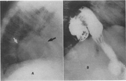
Figure 1: Hiatus
Hernia Visualized on a Roentgenogram of the Chest.
Certain lesions that formerly could not be diagnosed during life and therefore were considered of no significance are now known to be relatively common. The symptoms and signs of many of these conditions have been clarified, and the diagnosis can often be established clinically. An outstanding illustration of this development is afforded by hiatus hernia, which until a few years ago was a relatively unknown condition. The afflicted patients were incorrectly regarded as suffering from cholelithiasis, peptic ulcer, appendicitis and pancreatitis. Many gave a history of cholecystectomy, appendectomy, gastroenterostomy and lysis of adhesions, usually without symptomatic relief.
Because of the increasing numbers of cases uncovered in recent years, a symptom complex that is characteristic has emerged. The most typical complaint is a feeling of pressure or pain under the xiphoid during eating and relieved if the patient walks about for a few moments or takes a series of deep inhalations. The first swallows of food may be arrested at the level of the lower sternum, followed by a sense of "something passing through," after which the remainder of the meal can be finished in comfort.
Hiatus hernia may closely simulate organic heart disease because of chest pain and cancer because of hemorrhage, anemia or dysphagia. Statistical studies indicate that hiatus hernia occurs in 5 to 6 per cent of all persons. The condition may be diagnosable on roentgenograms of the chest and abdomen, being manifested by a soft-tissue density or a rounded area of increased radiance, often with an air-fluid level, in the retrocardiac region. Definitive diagnosis is made by the demonstration of a segment of the stomach above the diaphragm on x-ray examination utilizing an opaque meal. The x-ray has played a fundamental part in the establishment of this anomaly as an important entity.
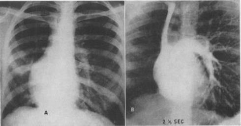
Another example of the conversion of a rare condition into one of great clinical interest is agenesis of a main pulmonary artery. Before 1954 only 11 cases of this developmental anomaly had been recorded, and the diagnosis had been established by surgery or autopsy in all. It can be identified on the roentgenogram of the chest, recognition being dependent on the presence of increased radiance in a lung that occupies less space than normal, deviation of the heart toward the ipsilateral side and absence of one main-pulmonary-artery shadow. Angiocardiography demonstrates the existence of a single pulmonary artery. The pathophysiologic significance of the anomaly lies in the fact that the affected lung does not participate in oxygen exchange because of deprivation of its pulmonary circulation. Its importance to the surgeon contemplating an aorticopulmonary anastomosis or pulmonary resection is obvious. Four cases have been diagnosed at the Boston City Hospital in the past two years. Increasing numbers will be uncovered as roentgenologists become more familiar with the condition:

Figure 2: Agenesis of
the Right Pulmonary Artery.
The dependability of roentgenographic diagnostic methods has emphasized the value of the x-ray in bringing about important medical advances. A striking example is afforded by the study of cases of acute massive bleeding from the upper gastrointestinal tract. Formerly, it was customary to defer attempts to establish the etiology by roentgenographic methods until the hemorrhage had ceased and the patient's condition had improved, in the mistaken belief that diagnostic procedures would increase the bleeding and endanger life.
In a series of over 1000 patients with acute, massive bleeding from the esophagus, stomach and duodenum, x-ray studies performed immediately after admission to the hospital established the correct diagnosis in practically all cases, and there were no complications because of the examinations. The practice of performing x-ray studies immediately after admission to the hospital of patients of this type has been adopted generally as a routine procedure and has saved many lives.
Limitations
Roentgenography has clearly defined limitations, and no implication that it is all-encompassing is intended. Many diseases cannot be diagnosed by this means. Some tend to place a greater reliance and dependence on the x-ray than its worth in comparison with other diagnostic procedures.
The changes demonstrable roentgenographically are macroscopic, and it is possible in many, but not in all, cases to deduce the exact microscopic picture of the underlying process from the gross alterations. The trained radiologist can visualize lesions as small as 4 or 5 mm. in diameter, although even larger ones may easily be missed.
The diagnosis of some diseases of the gastrointestinal tract continues to be uncertain, and negative findings in this region are less reliable than elsewhere in the body. This applies particularly to eosphageal varices, shallow gastric ulcers, early carcinoma, polyps and gastritis. In disease of the small bowel there usually must be marked stenosis or other serious derangement before diagnosis is possible.
Bronchitis, bronchopneumonia and bronchiectasis can exist for considerable periods and the clinical diagnosis can be clearly evident, and yet x-ray examination may reveal no abnormalities. In early miliary tuberculosis, pneumonitis and similar conditions, x-ray changes may be insignificant.
Osteomyelitis presents no roentgenographic changes until the third to the tenth day of the disease. In some cases fractures are not demonstrable by x-ray study for days after a trauma, particularly those of the flat bones such as the ribs, navicular and vertebras, most probably because the defect is so fine or lies at such an angle that it cannot be seen. Reexamination after the lapse of a few days shows slight displacement of the fragments or absorption along the line of cleavage that makes the break in continuity visible. In every patient with clinical evidence of a fracture, a negative examination should be repeated at short intervals.
Numerous lesions may produce practically identical x-ray changes. Diffuse pulmonary infiltrations may be due to congestion, fibrosis, scleroderma, sarcoidosis, yeast or fungous diseases, primary or metastatic neoplasm, bronchiectasis and virus pneumonia. A solitary density in the lung may occur in metastatic neoplasm, primary carcinoma, tuberculoma, encapsulated fluid, hamartoma, abscess, arteriovenous fistula and infarct. Miliary densities are seen in certain forms of pneumonia and tuberculosis, cancer, pneumoconiosis and hemosiderosis. Enlarged hilar shadows may be due to nonspecific infections, metastatic carcinoma, primary cancer of the lung, tuberculosis, sarcoidosis, lymphoma, pneumoconiosis, pneumonia, fungous diseases and certain congenital heart lesions.
The roentgenographic demonstration of a localized area of increased radiance in a bone presents a serious diagnostic problem. Differential diagnosis must include bone cyst, metastatic carcinoma, myeloma, osteomyelitis, sarcoma, lymphoma, neurofibromatosis, syphilis, fibrous dysplasia, eosinophilic granuloma, operative defect, nonosteogenic fibroma and giant-cell tumor. A zone of increased density in a bone may be due to an old fracture, sclerosing osteomyelitis, sarcoma, syphilis, osteoma, infarct, hypertrophic osteoarthropathy, metastatic carcinoma and osteitis deformans.
It is evident that roentgenographic methods of diagnosis have definite limitations and that negative findings do not always afford a complete sense of security. It is entirely proper for the clinician, under certain circumstances, to disregard negative or inconclusive x-ray studies.
Radiation Hazards
The fact that radiation has potentialities for harm is generally recognized and is particularly significant since the x-ray is used so widely in industry, research, science, the arts and warfare, as well as in medicine.
The deleterious effects that have been attributed to ionizing radiation are the following: leukemogenesis; carcinogenesis; shortening of the life span; and genetic disturbances.
Leukemogenesis
March made a survey of the mortality of doctors and stated that deaths from leukemia were nine times greater in radiologists than in other physicians. More careful analysis proved his figures in error, for the adjusted male death rate from leukemia indicated less than a twofold increase, and this is due in part at least to improved recognition of the disease, better methods of diagnosis and increased longevity.
The cases of leukemia have occurred largely in older members of the specialty, the disease apparently having been induced by excessive dosages administered during a prolonged period in an era when protective measures were less generally understood and often improperly used. Current studies indicate that the incidence of leukemia in radiologists is decreasing, whereas in other doctors utilizing x-rays it shows an increase. Despite the tremendous growth in the utilization of the x-ray, there has been only a moderate rise in the total number of cases.
Carcinogenesis
Most cases of carcinoma due to excessive exposure developed in the early years after Roentgen's discovery and involved the fingers and hands of those working with the rays, apparently because of failure to take proper precautions. In recent years radiation injury has occurred principally in surgeons and orthopedists who made it a practice to set fractures under the fluoroscope. Although the death rate from cancer was appreciably higher among British radiologists before 1921, this has not been true in more recent times since safe procedures came into general use.
Effect on Life Span
It has been reported that x-ray exposure shortens the life span and that radiologists die at an earlier age than their colleagues. There is no evidence that exposure to ionizing radiation over long periods has had any effect on the longevity of x-ray specialists. It is true that their average age at death has been lower than that of other physicians. This is due to a difference in age composition since radiology is a relatively young specialty and has not attracted the same variety and numbers of men as other fields of medicine. The death rate among 1377 British radiologists between 1897 and 1957 was no higher than that in the general population.
There has been a tremendous lengthening of the life span in the sixty-five years that have elapsed since the discovery of the x-ray. In 1900 the life expectancy at birth was forty-nine years; in 1959 it was sixty-nine years. Three generations of human beings have been subjected to ionizing radiation from medical x-ray studies and the fallout from nuclear testing. The actual lengthening of the life span has been due in part at least to the greater accuracy and promptness of diagnosis made possible by the x-ray.
Genetic Effects
Sterility, abortion, stillbirth and monstrosities are believed to ensue after excessive dosage of radiation to the gonads. Under natural conditions, mutations in the genes from radiation from cosmic rays, uranium, radium and artificial isotopes occur. Additional irradiation results in the acceleration of these changes rather than in the formation of new mutations. Animal experimentation indicates that the sequels of irradiation are largely deleterious so far as hereditary materials are concerned. Genetic implications are not clearly established in man, and it is generally accepted that genetic changes that may occur in animals do not necessarily have exact similarities in human beings.
Discussion
In recent years there has been a tremendous increase in the utilization of the x-ray. Orthopedists, urologists, neurosurgeons and others participate in fluoroscopic reductions of fractures, pyelography, myelography and similar procedures. There are 125,000 medical users of the x-ray in the United States, of whom about 50 per cent are dentists, 45 per cent practitioners and 5 per cent radiologists.
X-rays are not detected by the senses since they are invisible, odorless and silent and produce no sensation in their passage into and through the body. In many cases unsafe practices are continued until serious damage has resulted. Actual measurements of exposure received by participants in x-ray procedures have demonstrated that safety habits are poorly developed in the majority of nonqualified personnel.
The fluoroscope is frequently used by internists, pediatricians and other practitioners in their offices. The dangers to both patient and observer are particularly prevalent in this group. Reliance on the fluoroscope alone is neither dependable nor safe. Few practitioners devote the time required for a proper examination. Before the study, the eyes must be prepared by dark adaptation for twenty minutes. Lack of protective devices such as gloves, aprons and cones results in the delivery of large dosages of x-ray to the patient as well as the doctor. Many fluoroscopies being done by clinicians prove actually harmful, for lesions are missed. The nonradiologist is apt to transform a safe procedure when performed by the competent observer into one fraught with danger. The roentgenologist remains within safe exposure limits at all times, which often is not true of others.
The uninformed may become fearful because of overemphasis on the dangers and failure to understand how remote the possibility is that ill-effects will occur as the result of diagnostic roentgenographic examinations. It is generally agreed that all x-ray exposures should be reduced to the minimum, but it is neither desirable nor feasible to eliminate the x-ray from modern practice. Harmful results are avoided by the application of correct technics and proper safeguards. In the hands of trained personnel it is rarely if ever necessary to omit or postpone any roentgen examination that is indicated for the care of the patient. Careful measurements of dosages delivered during the performance of x-ray examinations prove that there is not even the remotest possibility that harm will result from properly performed roentgenographic studies. There is no evidence that the usual levels of diagnostic x-ray dosage are deleterious or that they cause cancer, leukemia, shortening of the life span or genetic damage.
The Roentgenologist
Specialism is now well established, and over a third of the doctors in the United States are specialists. Admittedly, disadvantages are associated with specialization, some of which cannot be eliminated. Despite the tendency to greater specialism, certain branches of medicine are of universal interest. Among these are psychiatry, allergy and radiology. No physician can practice successfully without at least a general knowledge of these fields.
The roentgenologist carries a special burden of responsibility because such great reliance and dependence are placed on his findings and conclusions. He acts as consultant and co-worker in the detection, analysis and integration of the data essential for the establishment of a diagnosis.
In certain obscure and occult lesions, the roentgenologist may be the one best able to demonstrate the abnormality. The x-ray studies do not always establish the genesis, nature and course of the deviations demonstrable roentgenographically. However, by bringing the findings to the attention of the clinician, pathologist and others such studies may make the condition more clearly understood through the combined efforts of the various members of the group.
The highest standards of excellence should prevail in every x-ray study. Everyone who undertakes the interpretation of roentgenographic findings must remain completely objective and not lose the unbiased judgment essential in medicine.
Teaching
The x-ray has become virtually indispensable in teaching since it demonstrates graphically both normal states and pathologic conditions. Of equal importance is the fact that roentgenographic studies have proved that there are fundamental and highly important variations from the previous teachings in numerous areas of medicine. The older concepts were based in large part on observations made in the adynamic viscus during dissection or on the autopsy table and at the time of operation. Roentgenography presents the truer picture because it causes little or no interference with the existing patterns and relations.
There are of necessity different types of instruction for those specializing in radiology than for the undergraduate and graduate student. The hospital resident requires four years of training, observation and experience to be considered qualified for certification in the specialty. The sequels of the incorrect administration of a drug or the faulty performance of a surgical procedure are so prompt and dramatic that few are willing to accept the consequences unless adequately prepared and competent. The harmful potentialities of an incorrect or missed x-ray diagnosis may be less obvious, and their ill-effects not so apparent and direct. Nevertheless, they are no less serious and are equally to be avoided and condemned.
Teaching of roentgenology to nonradiologists comprises indications, capabilities and hazards, with theoretical discussions and general descriptions of technics and practical methods. The purpose of the instruction is to provide a basic knowledge of diagnostic criteria to enable the physician to distinguish between good and bad practices.
Future
In the early days, the x-ray served chiefly in the diagnosis of fractures, the demonstration of metallic foreign bodies and the detection of opaque calculi. Further applications developed rapidly, and roentgenology from its very inception proved an expanding field.
Until recent years the conditions diagnosable by x-ray study for the most part were static. There is in process of development a broader approach, termed the dynamic phase. Technical advances by the physicist, engineer and research scientist complement and extend the work of the roentgenologist. The potentialities of future growth are vast and there is no discernible limit to the progress which may be expected ultimately.
Equipment
In the period immediately after Roentgen's discovery, a radiograph required many minutes' exposure time. Today roentgenograms can be taken in a thousandth of a second. It may soon be possible to produce x-rays without bulky transformers and control stands. United States Army physicians are perfecting an x-ray machine that employs a radioactive isotope as a source of current. New types of x-ray tubes are being developed. One of the advances envisioned is a tube that will be discarded after a single exposure, being similar to the flash bulb used in photography.
There is continuous improvement in the speed of x-ray films and intensifying screens, greatly lessening the dosages delivered during the making of roentgenograms. At present, three or more separate liquids are necessary to process a film. These will be replaced by a single solution, which will function more rapidly and with greater efficiency. Automatic devices eliminate the handling of wet films. The roentgenogram can be developed, washed and dried in five minutes. Automatic units are costly and are practical only in large medical institutions but soon will be available for all hospitals and private offices.
Television
The transmission of roentgenograms by television permits areas lacking the services of a trained roentgenologist to obtain an interpretation with a minimum of delay. There is also in process of development a recording device that will permit high-speed televising of x-ray films, eliminating the delays entailed in processing. Immediately after the recording appears on the drum face, the image can be reproduced and made available for study by means of a television tube.
Image Intensification
The present intensifiers accentuate the x-ray effect on the fluoroscopic screen by a factor of approximately 1500. This results in a brighter image that requires only a fraction of the exposure entailed in conventional fluoroscopy. It will soon be possible to obtain intensifications of 5000 times or more. When combined with television apparatus, the roentgenoscopic image can be projected at a distance and viewed simultaneously by multiple observers.
The amplifiers are limited in size, the field of vision being only a circle 13 cm. in diameter. There are in process of development devices measuring 30 cm. or more.
Cineradiography
In cineradiography the fluoroscopic image is recorded permanently on motion-picture film while being observed by the fluoroscopist. This has tremendous diagnostic potential and eventually will supplant current roentgenoscopic procedures since it permits the recording of dynamic phenomena. The cinefilmed sequences have the advantage of being easily filed and readily available for study and restudy.
Body-Section Radiography
Body-section radiography is a type of x-ray examination in which predetermined layers of the body are visualized, with exclusion of other areas above or below the segment under observation. It entails the taking of a series of roentgenograms at varying depths, usually at distances 5.0 to 10.0 mm. apart. The procedure has been termed laminography, tomography, planigraphy and roentgen section. It results in the delivery of considerable amounts of radiation to the parts under observation. The development of the book type of cassette, which affords a series of 7 roentgenograms with a single exposure, has resulted in a great saving of time and reduces the radiation to a practically negligible level. Further improvement will make body-section radiography more widely applicable.
Xeroradiography
Xeroradiography obviates the need for x-ray film. Electrostatically charged plates of aluminum coated with photoconductive selenium are exposed to x-rays, and a radiograph is produced within a minute or less. It requires no chemical solutions or darkroom.
The recording medium cannot be rendered useless by the ionizing radiation of an atomic bombing or radioactive fallout since the plates are insensitive to x-rays until electrically activated. It has an important place during disasters such as earthquakes, floods and hurricanes because it does not require fresh water. It is not ideal for the study of the heavier parts of the body. Further refinement will make it generally applicable in medical radiography.
Research
Active investigation utilizing x-rays began almost immediately after the announcement of Roentgen's discovery. Scientists have continued to find x-ray study invaluable in probing the structure and function of the body.
Improved contrast mediums are essential for the expansion of x-ray diagnostic procedures. A significant development is an organic iodide solution that can be used in place of barium sulfate when retention in the intestines, body cavities or fistulous tracts is anticipated. It is of value in the diagnosis of tracheoesophageal and gastrointestinal fistulas, obstructions and perforations, although the degree of contrast obtained with the water-soluble mediums is limited.
Research is being directed toward improvement in the roentgenographic visualization of the cerebral ventricles, the spinal canal and the nerve roots. Air, iodized oil, iophendylate and other mediums in use today are not ideal. Substances to opacify the viscera, gonads and lymphatics without producing undesirable side effects are being sought in many laboratories.
Angiography
Angiography clarifies many previously obscure points in the study of the anatomy, physiology and dynamics of the circulation, as well as being of great value as a diagnostic tool. No one would have believed a generation ago that it would actually be possible to see the circulation within the heart, lungs, brain, kidneys and extremities and that vascular lesions, which previously could be diagnosed only at autopsy, would be demonstrable with clarity and ease.

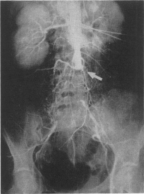
Figure 3: Aortogram
Showing Occlusion of the Aorta.
The injection of opaque material through a catheter inserted into the brachial or femoral artery and passed into the aorta makes it possible to visualize the coronary, carotid, renal and other arteries and to determine whether the vessels are narrowed, occluded, tortuous or otherwise abnormal. The procedure is being refined, and still wider applications of its use will be uncovered.
Automation
Automation in x-ray diagnosis is in process of development. In his practice, the radiologist analyzes the available data, considers every condition that might produce the changes and determines what he believes to be the correct or most probable diagnosis. Only lesions with which he is familiar come into consideration, and this is dependent on the breadth of his knowledge. The most significant disease in the case under study may not occur to him. An automatic device will consider all possible lesions every time it is put into operation.
Scanning devices to record variations in densities on the roentgenogram are finding a limited field of usefulness. Units of this type will eventually occupy a place in medical practice. It is not inconceivable that a robot will take the x-ray film, process it and feed it into an analyzer, which will immediately produce the diagnosis. Although no mechanical device ever can replace the skill, experience and diagnostic ability of the trained and competent radiologist, automation will have a very important place in diagnostic roentgenology in the future.
_______________
Ritvo, Max. "The Role of Diagnostic Roentgenology in Medicine." New England Journal of Medicine 262, no. 24 (1960): 1201-09.
View all Annual Orations
